Newsletter 05-2017
18th May 2017
Scannexus in the media
Are you curious about what is going on at our facilities? We would like to give you a small impression.

"Galileo"
The creators of the RTL5 Television Program Galileo visited us for filming an interesting study;
Jorn's body is controlled by someone else: scientists at the University of Maastricht can move other people with their own brain. And is it possible to read somebody's mind?
Check out this item and see what can be achieved with imaging today!

"Sophie in de Mentale Kreukels"
Sophie in de Mentale Kreukels (mental cracks) did a part of her research at our facilities for her TV program at BNN.
Sophie herself got a burnout four-and-a-half years ago. In the Netherlands, more than one million people a year suffer from these symptoms. What causes this problem to grow bigger, and why is it starting at younger ages? What should and can we do to prevent it? In each episode, Sophie tries different alternative therapies.
'Click here' to see the episode in which she visited our facilities.

Introduction Kim Brouwers, MRI Operations & Support Technician
It’s been nearly 15 years ago since I started working with an MRI scanner as a technologist for the first time. I think the opportunities these systems accomplish are very interesting and changing all the time. Observing how new developments influence patient care and treatment in the clinic, expanded my own MRI interests from daily care to the research aspect of the modality as well. Scannexus plays an important role in these developments, functioning as a bridge between patient care and new technology.
Next to Scannexus, I am also working at the department of radiology at the MUMC (Maastricht University Medical Centre). My main interest is cardiac imaging, both on regular MRI systems as on the PET-MRI (Positron Emission Tomography - Magnetic Resonance Imaging). I have been working with a PET-MRI system for three years now. The PET component made me more aware of all the different metabolic processes in human body. These processes are very interesting and I therefore started working for NUTRIM several hours a week. Within this research project MR spectroscopy is used a lot and it is another way of metabolic imaging.
High field imaging fits very well in all of this I think. I am looking forward to the opportunity to learn new techniques and work with state-of-the-art systems and new colleagues. Not only in brain related research but also within other parts of the body, as the campus is going to expand the research- and business possibilities to a broader range of the body.

Introduction Bente Faarts, MRI Operations & Support Technician in training
I'm always up for a new challenge. And now I'm here, at Scannexus. I am also working at the department of radiology at MUMC (Maastricht University Medical Centre) as a Regular Radiographer and wanted to specialize my experience in the MRI field. Since 2015, after graduating from MBRT (Medical Imaging/Radiation Oncology), I have been working at MUMC. Within this area I want to further expand my knowledge and experience as an Operations & Support Technician.
September this year I will return to college to complete the specialization module ‘Magnetic Resonance’. Hopefully, next year I will be a fully trained member of the Scannexus Support Team. In the meantime, I will obtain a lot of knowledge from my colleagues and I will try to do my best by helping where I can.
My ultimate goal is to become an Operations & Support Technician within Scannexus to combine this with my function as a Regular Radiographer within MUMC.
MRI is the future. Scannexus provides a unique, integrated platform that combines state-of-the art technology with know-how (and access to patients) and internationally renowned expertise in post-acquisition processing of data in relation to Ultra-High-Field human MRI. Therefore, I'm grateful for getting the opportunity to play my part in the promising future of this medical research area.
Publications from our employees
Comparing UHF MRI to historical anatomical atlases
Research using Ultra High Field MRI, such as those available at Scannexus, continues to reveal insights into the human body, especially the brain. A recent publication in the British Journal of Radiography by our team member Christopher Wiggins PhD (in collaboration with the Max Planck Institute for Human Cognitive and Brain Sciences in Leipzig, Germany and CEA/NeuroSpin in Saclay, France) shows some of the unique power of these scanners.
In this example, the Corpus Callosum - the main nerve fiber tract that connects the left and right hemispheres of the brain - is shown to have a structure composed of parallel ribbons. This paper compares 7T in-vivo images of the Corpus Callosum to historical anatomical atlases going back 200 years, and shows that UHF MRI is capable of seeing in-vivo what has previously only been seen! in ex-vivo dissection. The ability to reveal such fine structures in-vivo will aid the study of the brain, especially in regards to longitudinal studies of brain development and comparison across individuals.
'Click here' to read the complete article.
Development of visual category selectivity in ventral visual cortex does not require visual experience
In a collaboration with the Catholic University of Leuven, Job van den Hurk authored a paper that appeared this week in PNAS:
The brain’s ability to recognize visual categories is guided by a category-selective brain region, called Ventral-Temporal Cortex (VTC). Whether visual experience is required for the functional organization of VTC into distinct functional subregions remains unknown, hampering our understanding of the mechanisms that drive category recognition.
This paper demonstrates that VTC in individuals who were blind since birth shows robust discriminatory responses to natural sounds representing different categories (faces, scenes, body parts, and objects). These activity patterns in the blind also could predict successfully which category was visually perceived! by controls. The functional cortical layout in blind individuals showed remarkable similarity to the well-documented layout observed in sighted controls, suggesting that visual functional brain organization does not rely on visual input.
'Click here' to read the complete article.
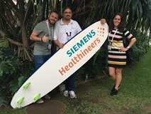
Scannexus at ISMRM 2017
A remarkable number of white legs were spotted on the sunny shores of Honolulu at the end of April. MRI-aficionado’s from all over the world gathered at the beautiful Hawaiian island Oahu for the ISMRM (International Society for Magnetic Resonance in Medicine) 2017 conference.
For almost a full week, the 25th edition of this conference was packed with educational courses, scientific sessions, poster presentations and business exhibitions. Thousands of scientists and entrepreneurs intermingled to share knowledge, to gain ideas and to get updated on the latest fashion for scientists (the traditional socks-in-sandals are out of fashion, it turns out)!
Nadine, Chris and Job attended ISMRM on behalf of Scannexus. They spoke with several research groups and companies, worked out international collaborations, and learned a lot about hardware, software and applications for neuro-, spinal-, cardiac- and MSK-MRI. In addition, Chris delivered two interesting and light-hearted presentations. Luckily, there was no time for us to miss Maastricht, as our city was very well represented at ISMRM (no less than 25 times!). Excellent researchers from our Faculties Psychology and Neuroscience (FPN) and Medicine, Health and Lifesciences (FHML) as well as the MUMC+ presented their research with flair and dedication.
For the upcoming months, the inspiration and energy we got from ISMRM will bring us further in our journey to innovate and be fascinated. See you next year in Paris!
< Back to overview News